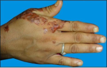
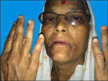
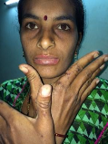
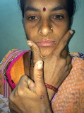
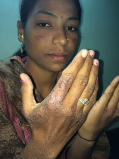
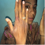
Introduction: Anaemia is defined as a decrease in the total amount of RBCs or haemoglobin in the blood. Anaemia patients show skin signs like pallor, koilonychias, and hair loss. Aim: The aim of the study is to find the cause of typical rash and confirm the diagnosis. Materials/Methods: This retrospective study involves collection of 26 patients showing typical skin lesions. All subjects involved in this study are categorized according to age, sex, type of lesions and site involved. All were investigated for hematological parameters mainly haemoglobin (Hb), mean corpuscular volume (MCV), mean corpuscular haemoglobin (MCH). Everyone was showing reduced parameters with microcytic hypochromic red blood cells suggesting iron deficiency anaemia. These patients were further investigated for other causes of anaemia and diagnosis was confirmed by serum iron levels, total iron binding capacity, serum ferritin and transferrin saturation. Other causes like zinc deficiency, pellagra, contact dermatitis were also ruled out. Results: Almost all patients are females except two males. Majority of them are young females. The skin lesions can present isolated on face or hands or both sites. Each and every subject is confirmed to have iron deficiency anemia. Limitations: This is a single center study. Conclusion: These typical eczematous hand and perioral lesions were highlighting the importance of looking for iron deficiency in affected patients without a more obvious cause.
| Published in | International Journal of Clinical Dermatology (Volume 9, Issue 1) |
| DOI | 10.11648/j.ijcd.20260901.15 |
| Page(s) | 34-39 |
| Creative Commons |
This is an Open Access article, distributed under the terms of the Creative Commons Attribution 4.0 International License (http://creativecommons.org/licenses/by/4.0/), which permits unrestricted use, distribution and reproduction in any medium or format, provided the original work is properly cited. |
| Copyright |
Copyright © The Author(s), 2026. Published by Science Publishing Group |
Iron Deficiency Anaemia, Eczema, Cutaneous Marker
Index | HB gm% | RBC 3.8-5.3 | WBC | HCT 36-56 | MCV80-100 | MCH27-32 | MCHC32-36 | PLT120-380 | RDW10-16 |
|---|---|---|---|---|---|---|---|---|---|
1 | 6.8 | 4.3 | 23.2 | 59.8 | 17.5 | 29.3 | 1.65 | ||
2 | 8.3 | 4.16 | 6.5 | 24.1 | 57.9 | 20 | 34.4 | 333 | 16.6 |
3 | 9.3 | 4.69 | 8.5 | 27.1 | 57.8 | 19.8 | 34.3 | 329 | 15.7 |
4 | 6.1 | 4.22 | 7.5 | 20.6 | 48.8 | 14.5 | 29.6 | 357 | 16.8 |
5 | 6.3 | 4.17 | 5 | 21.4 | 51.3 | 15.1 | 29.4 | 304 | 17.1 |
6 | 7.3 | 3.84 | 9.7 | 23 | 59.9 | 19 | 31.7 | 352 | 16 |
7 | 5.7 | 3.84 | 6.3 | 20.5 | 53.4 | 14.8 | 27.8 | 453 | 18.3 |
8 | 5.9 | 4.42 | 9.3 | 20.7 | 46.8 | 13.3 | 28.5 | 414 | 18 |
9 | 8 | 4.39 | 4.8 | 25.1 | 57.2 | 18.2 | 31.9 | 425 | 15.5 |
10 | 6.1 | 4.02 | 7.2 | 20.1 | 50 | 15.2 | 30.3 | 279 | 20.2 |
11 | 6.6 | 4.7 | 6 | 20.4 | 50.1 | 16.2 | 32.4 | 491 | 17.2 |
12 | 7.7 | 4.14 | 6.1 | 22.9 | 53.3 | 18.6 | 33.6 | 360 | 17.5 |
13 | 8.6 | 4.37 | 8.9 | 24.1 | 55.1 | 19.5 | 35.3 | 474 | 16.3 |
14 | 7.2 | 3.79 | 7.2 | 21.8 | 57.5 | 18.7 | 32.6 | 359 | 15.6 |
15 | 5.6 | 4.04 | 8.8 | 21.8 | 54 | 13.9 | 25.7 | 390 | 16.8 |
16 | 8.1 | 4.7 | 6.4 | 26.6 | 56.6 | 17.2 | 30.5 | 138 | 16.8 |
17 | 7.7 | 5.07 | 6.6 | 27.2 | 53.6 | 15.2 | 28.3 | 329 | 16.3 |
18 | 4.3 | 3.23 | 3.1 | 18.1 | 56 | 13.3 | 23.8 | 409 | 15.7 |
19 | 6.5 | 4.16 | 4 | 23.4 | 56 | 15.7 | 27.8 | 325 | 18.7 |
20 | 3.7 | 2.64 | 7.7 | 24.1 | 53 | 13.9 | 26 | 361 | 18.5 |
21 | 6.4 | 4.85 | 12.5 | 21.9 | 45.3 | 13.1 | 29.2 | 344 | 24 |
22 | 5.3 | 4.1 | 7 | 20.9 | 50.9 | 12.9 | 25.4 | 394 | 20.4 |
23 | 6.5 | 5.28 | 12700 | 23.7 | 44.9 | 12.3 | 27.4 | 443 | 20.8 |
24 | 8.3 | 3.95 | 7100 | 24.2 | 61.1 | 20.9 | 34.3 | 399 | |
25 | 6.5 | 4.29 | 4400 | 27.4 | 63.87 | 15.15 | 23.72 | 429 | 19.3 |
26 | 7.4 | 4.69 | 9100 | 27.6 | 58.8 | 15.8 | 26.8 | 262 | 14.2 |
Index | PCT.1-1 | MPV5-10 | PDW12-18 | sr iron60-150 | TIBC250-400 | SR FERRITIN15-230 | TRANSFERRIN SATURATION20-50% |
|---|---|---|---|---|---|---|---|
1 | |||||||
2 | 0.18 | 5.4 | 15.5 | ||||
3 | 0.14 | 4.3 | 16.4 | ||||
4 | 0.21 | 5.9 | 15.1 | ||||
5 | 0.18 | 6 | 15.9 | 15 | 466 | 2.9 | 3 |
6 | 0.17 | 4.8 | 17.3 | ||||
7 | 0.2 | 4.5 | 15 | ||||
8 | 0.22 | 5.4 | 14.8 | 32 | 352 | 1.02 | 9.09 |
9 | 0.22 | 5.2 | 17.4 | 15 | 502 | 2.4 | 3 |
10 | 0.18 | 6.3 | 15.2 | 13.8 | 579.73 | 2.7 | 2.38 |
11 | 0.22 | 4.4 | 16.5 | ||||
12 | 0.13 | 3.6 | 17.5 | ||||
13 | 0.24 | 5 | 16.7 | ||||
14 | 0.13 | 3.5 | 19 | ||||
15 | 0.2 | 5 | 14.7 | ||||
16 | o.11 | 5.7 | 15.1 | ||||
17 | 0.14 | ||||||
18 | 0.4 | 10 | 25 | ||||
19 | 0.325 | 8.6 | 17.4 | ||||
20 | 0.278 | 7.7 | 14 | ||||
21 | 0.25 | 10.8 | |||||
22 | 0.32 | 8.2 | 15.4 | ||||
23 | 0.39 | 8.8 | 13.6 | ||||
24 | |||||||
25 | zinc -178.9 (54-151) | ||||||
26 | |||||||
MARKERS | IRON DEFICIENCY ANAEMIA | B-THALASEMIA | CHRONIC CAUSE |
|---|---|---|---|
MCV/RBC | >13 | <13 | |
RDW | INCREASED | NORMAL | |
SR IRON | DECREASED | NORMAL | DECREASED |
FERRITIN | DECREASED | NORMAL | INCREASED NORMAL |
FEP | INCREASED | NORMAL | |
MCHC | DECREASED | ||
PLATELET COUNT | INCREASED | ||
TARGET CELLS | ABSENT | PRESENT | |
TIBC | INCREASED | NORMAL | DECREASED |
THERAPEUTIC TEST WITH ORAL IRON | RISE IN HB | NO RISE IN HB | NO RISE |
Vit | Vitamin |
| [1] | "Anemia: Practice Essentials, Pathophysiology, Etiology". November 9, 2021. Retrieved February 8, 2022. |
| [2] | Haematology: clinical principles and applications (3rd ed). Philadeiphia: Saunders. p. 220. |
| [3] | Textbook of Dermatology. Ed Rook A, Wilkinson DS, Ebling FJB. Fourth edition. |
| [4] | Canadian Medical Association Journal. 119(8): 884-886. |
| [5] | Sleep Medicine. 8(3): 247-51. |
| [6] | ”How is iron deficiency anaemia is treated”. NHLBI. 26 March 2014. |
| [7] | Goldman L, Schafer A (2016). Goldman-Cecil Medicine. pp. 1052-1059, 1068-1073. |
| [8] | Henry’s Clinical Diagnosis and Management by Laboratory Methods. pp. 84-101, 559-605. |
| [9] | Jake Turner; Meghana Parsi; Madhu Badireddy. Last Update: Anemia. August 8, 2023. |
| [10] | Usuki K. [Anemia: From Basic Knowledge to Up-to-Date Treatment. Topic: IV. Hemolytic anemia: Diagnosis and treatment]. Nihon Naika Gakkai Zasshi. 2015 Jul 10; 104(7): 1389-96. |
| [11] | O'Farrill-Santoscoy F, O'Farrill-Cadena M, Fragoso-Morales LE. [Evaluation of treatment of iron deficiency anemia in pregnancy]. Ginecol Obstet Mex. 2013 Jul; 81(7): 377-81. |
| [12] | Rashid A. A 65-year-old man with anemia: diagnosis with peripheral blood smear. Blood Res. 2015 Sep; 50(3): 129. |
| [13] | Bottomley SS, Fleming MD. Sideroblastic anemia: diagnosis and management. Hematol Oncol Clin North Am. 2014 Aug; 28(4): 653-70, v. |
| [14] | Association of Zinc with Anemia. Sen-Shyong Jeng, Yen-Hua Chen. 2022 Nov 20; 14(22): 4918. |
| [15] | Huck, J. the blood picture of uncomplicated pellagra with a review of the literature. Bull. Jons Hopkins Hosp., 1923, 34, 157. |
| [16] | Turner, R. H., with the assistance of E. Shelton, Ethrocytes in pellagra. Am. J. M. Sc., 1933, 185, 381. |
APA Style
Balkrishna, B. A. (2026). The Classic Dermatological Signs of Iron Deficiency: A Case-Series. International Journal of Clinical Dermatology, 9(1), 34-39. https://doi.org/10.11648/j.ijcd.20260901.15
ACS Style
Balkrishna, B. A. The Classic Dermatological Signs of Iron Deficiency: A Case-Series. Int. J. Clin. Dermatol. 2026, 9(1), 34-39. doi: 10.11648/j.ijcd.20260901.15
@article{10.11648/j.ijcd.20260901.15,
  author = {Bhokare Anil Balkrishna},
  title = {The Classic Dermatological Signs of Iron Deficiency:
A Case-Series},
  journal = {International Journal of Clinical Dermatology},
  volume = {9},
  number = {1},
  pages = {34-39},
  doi = {10.11648/j.ijcd.20260901.15},
  url = {https://doi.org/10.11648/j.ijcd.20260901.15},
  eprint = {https://article.sciencepublishinggroup.com/pdf/10.11648.j.ijcd.20260901.15},
  abstract = {Introduction: Anaemia is defined as a decrease in the total amount of RBCs or haemoglobin in the blood. Anaemia patients show skin signs like pallor, koilonychias, and hair loss. Aim: The aim of the study is to find the cause of typical rash and confirm the diagnosis. Materials/Methods: This retrospective study involves collection of 26 patients showing typical skin lesions. All subjects involved in this study are categorized according to age, sex, type of lesions and site involved. All were investigated for hematological parameters mainly haemoglobin (Hb), mean corpuscular volume (MCV), mean corpuscular haemoglobin (MCH). Everyone was showing reduced parameters with microcytic hypochromic red blood cells suggesting iron deficiency anaemia. These patients were further investigated for other causes of anaemia and diagnosis was confirmed by serum iron levels, total iron binding capacity, serum ferritin and transferrin saturation. Other causes like zinc deficiency, pellagra, contact dermatitis were also ruled out. Results: Almost all patients are females except two males. Majority of them are young females. The skin lesions can present isolated on face or hands or both sites. Each and every subject is confirmed to have iron deficiency anemia. Limitations: This is a single center study. Conclusion: These typical eczematous hand and perioral lesions were highlighting the importance of looking for iron deficiency in affected patients without a more obvious cause.},
 year = {2026}
}
TY  - JOUR
T1  - The Classic Dermatological Signs of Iron Deficiency:
A Case-Series
AU  - Bhokare Anil Balkrishna
Y1  - 2026/03/09
PY  - 2026
N1  - https://doi.org/10.11648/j.ijcd.20260901.15
DO  - 10.11648/j.ijcd.20260901.15
T2  - International Journal of Clinical Dermatology
JF  - International Journal of Clinical Dermatology
JO  - International Journal of Clinical Dermatology
SP  - 34
EP  - 39
PB  - Science Publishing Group
SN  - 2995-1305
UR  - https://doi.org/10.11648/j.ijcd.20260901.15
AB  - Introduction: Anaemia is defined as a decrease in the total amount of RBCs or haemoglobin in the blood. Anaemia patients show skin signs like pallor, koilonychias, and hair loss. Aim: The aim of the study is to find the cause of typical rash and confirm the diagnosis. Materials/Methods: This retrospective study involves collection of 26 patients showing typical skin lesions. All subjects involved in this study are categorized according to age, sex, type of lesions and site involved. All were investigated for hematological parameters mainly haemoglobin (Hb), mean corpuscular volume (MCV), mean corpuscular haemoglobin (MCH). Everyone was showing reduced parameters with microcytic hypochromic red blood cells suggesting iron deficiency anaemia. These patients were further investigated for other causes of anaemia and diagnosis was confirmed by serum iron levels, total iron binding capacity, serum ferritin and transferrin saturation. Other causes like zinc deficiency, pellagra, contact dermatitis were also ruled out. Results: Almost all patients are females except two males. Majority of them are young females. The skin lesions can present isolated on face or hands or both sites. Each and every subject is confirmed to have iron deficiency anemia. Limitations: This is a single center study. Conclusion: These typical eczematous hand and perioral lesions were highlighting the importance of looking for iron deficiency in affected patients without a more obvious cause.
VL  - 9
IS  - 1
ER  -
Apex Skin & Laser Center, Malegaon, India